
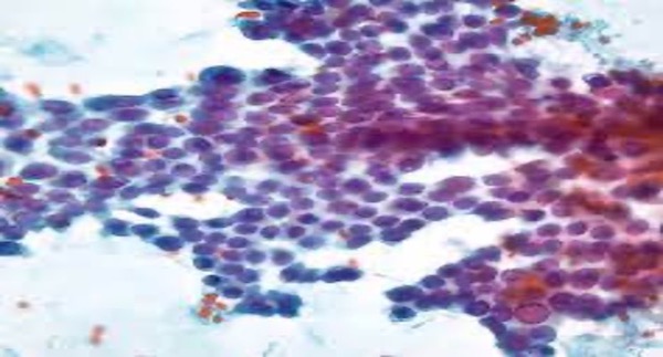
Cytology, or cytopathology, is currently known as a subspecialty of pathology. While histopathology evaluates tissue, cytopathology evaluates cells.
The material obtained may sometimes be cells spontaneously shed into the normal anatomical cavities of the body, or sometimes cells aspirated from the relevant organ or anatomical region with the help of a syringe.
It is one of the preferred methods because it is cheap, easy, and causes minimal damage to humans or tissue.
It may not always allow for ideal diagnosis or pathological examination. In recent years, tissue images can be obtained using the cell block method from good aspiration material.
The cell block method is a routine procedure performed on all cytologic specimens submitted to our laboratory. If a thorough aspiration is performed, further processing with the cell block method is possible, providing optimal support for patient diagnosis, treatment, and follow-up.
body fluids
It is the process of aspirating fluids from anatomical regions within the human body for pathological examination. Examples include the abdominal cavity, thoracic cavity, and bronchial fluid.
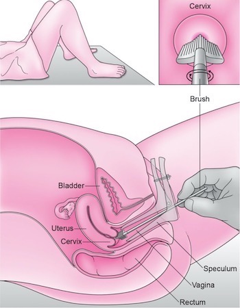
Cervical Cytology (Smear)
Cytology is one of the most frequently used methods. Cells shed from the cervix are collected using a special brush and transferred to a slide. They are then stained with special dyes and examined. A Pap smear is a screening test, meaning it's performed not only on patients with suspected or suspected disease but also on healthy individuals.
It is used to detect cellular disorders, precancerous cells and infections in the cervix, especially cervical cancer.
This way, cellular disorders can be detected at an early stage before they develop into uterine and cervical cancer, allowing the patient to fully recover.
Fine Needle Aspiration Cytology (FNAC)
This practical, minimally invasive, rapid, and cost-effective method is used to diagnose certain diseases. It relies on the examination of cells in fluid obtained from the thyroid, breast, lung, and many other organs through a needle. This method aims to diagnose diseases. While it may not always allow for ideal diagnosis or pathological examination, in recent years, tissue images can be obtained using the cell block method from well-aspirated specimens. The cell block method is a routine procedure performed on every cytologic specimen submitted to our laboratory. If aspiration is performed properly, further processing with the cell block method is possible, providing optimal support for the diagnosis, treatment, and follow-up of the patient.
Phone +90(216) 469 14 68
Mobile+90(535) 977 49 13
E-mail: info@marmarapatoloji.com

